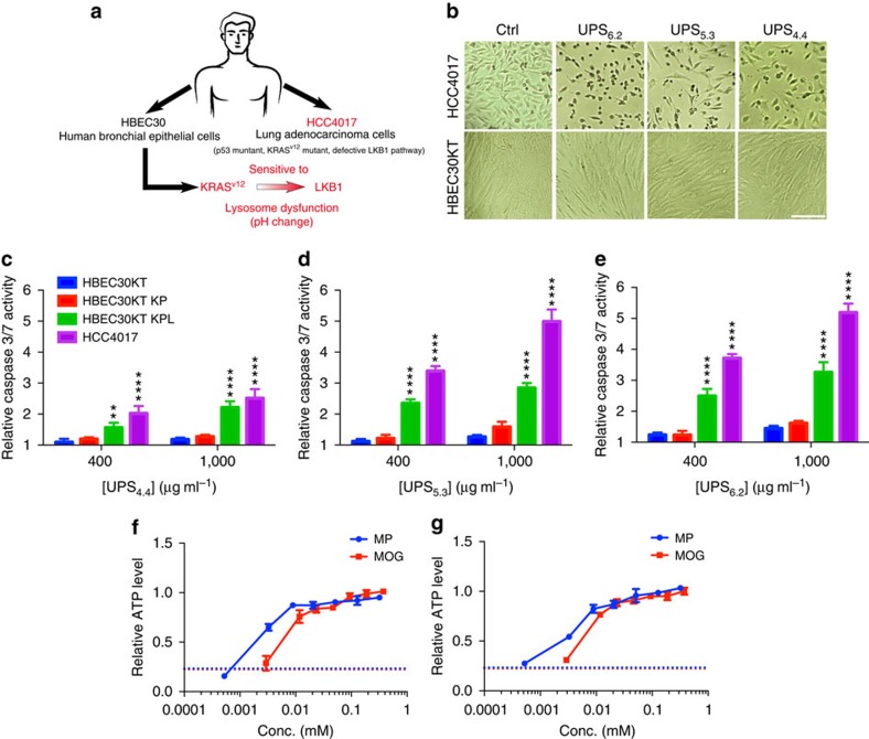
Figure 6. UPS nanoparticles selectively kill NSCLC cells that are sensitive to lysosomal stress.
(a) Schematic of the cell models employed and their corresponding vulnerabilities to lysosomal maturation. (b) Bright field images indicating the relative viability of HBEC30 KT and HCC4017 cells with and without exposure to UPS at effective doses (UPS6.2 and UPS5.3=400 μg ml−1, UPS4.4=1,000 μg ml−1). Scale bar, 100 μm. (c–e) Caspase3/7 activity in HBEC30KT, HBEC30KT KP, HBEC30KT KPL and HCC4017 cells was measured 72 h after exposure to the indicated doses of UPS. Two-way analysis of variance and Sidak's multiple comparison tests were performed to assess statistical significance of observed differences between HBEC30KT and HCC4017, and HBEC30KT KP and HBEC30KT KPL, α=0.05, **P<0.01, ****P<0.0001. (f,g) Cellular ATP levels were measured after exposure of HCC4017 (f) and HBEC30 KT KPL (g) to 1,000 μg ml−1 UPS6.2 for 72 h together with the indicated concentrations of methyl pyruvate (MP), dimethyl-2-oxoglutarate (MOG) or water (dash line). Values were normalized to no treatment (that is, without UPS) controls. Error bars indicate s.d., n=4.

