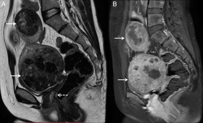
Figure 1.
(A) Sagittal T2-weighted and (B) sagittal postcontrast T1-weighted MRI showing two heterogeneously enhancing circumscribed masses (white arrows) in the pelvis and hypogastrium, which appear hypointense to muscle. The mass in the pelvis is indenting on the dome of the urinary bladder with preserved fat planes. No obvious uterine tissue is seen between the rectum and bladder (dashed white arrow).

