
Fig. 4.
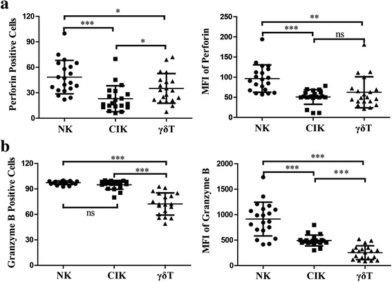
Perforin and granzyme B production in expanded NK, CIK, and γδ T cells. Three types of immune cells from twenty cancer patients were harvested after 14 days induction in vitro. NK and CIK cells were stained with mAbs to CD3 and CD56, and γδ T cells were stained with CD3 and Vγ9. After fixation and permeabilization, cells were stained for perforin and granzyme B using specific conjugated anti-human cytokine mAbs. a Perforin-positive cells and mean fluorescence intensity (MFI) of NK, CIK, and γδ T cells. b Granzyme B-positive cells and MFI of NK, CIK, and γδ T cells. Results are expressed as mean ± SD, n = 20. * p < 0.05, ** 0.001 < p < 0.01, *** p < 0.001, ns p > 0.05