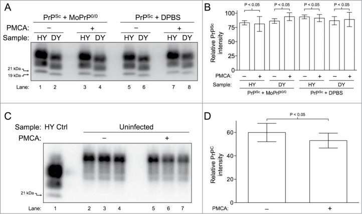
Figure 3.

Clearance of PrP is not supported by PMCA. (A) Western blot of HY TME and DY TME showing an absence of PrPSc clearance during PMCA. HY TME and DY TME diluted in MoPrP0/0 brain homogenate without sonication (Lanes 1–2); HY TME and DY TME diluted in MoPrP0/0 brain homogenate subjected to PMCA (Lanes 3–4); HY TME and DY TME diluted in DPBS without sonication (Lanes 5–6); and HY TME and DY TME diluted in DPBS subjected to PMCA (Lanes 7–8). (B) Bar graph comparing the relative intensity of each sample before and after sonication (n = 4 per experimental group). (C) Western blot of uninfected hamster brain homogenate showing the absence of PrPC clearance during PMCA. Uninfected brain homogenate without PMCA (Lanes 2–4) and after PMCA (Lanes 5–7). (D) The relative average intensities of PrPC as quantified from the Western blot analysis of each sample before and after PMCA (n = 3 per experimental group). The migration of the 19 or 21 kDa unglycosylated PrPSc polypeptides is indicated on the left of panels A and C.
