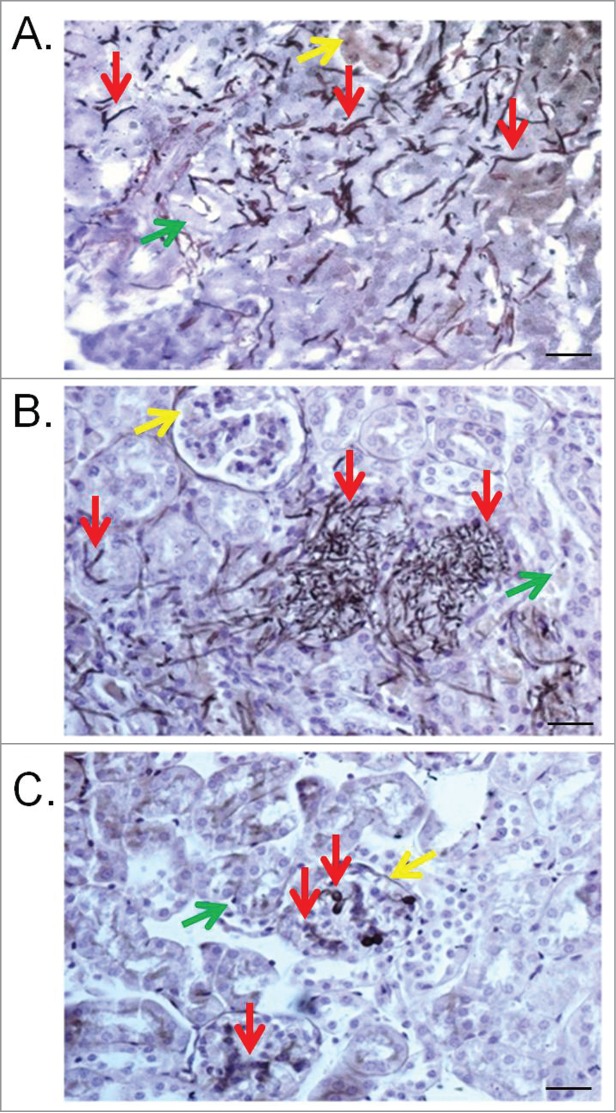
Figure 6.

Kidney lesions in the murine model caused by T. asahii 07 (A), T. asteroides 01 (B) or T. inkin (C) infections 6 days after challenge. Kidney transversal sections showing hyphae, blastoconidia and arthroconidia (red arrows) in renal tubules (green arrows) and some of them in the glomerular structure (yellow arrows). GMS and hematoxylin stain. Bars, 20 μm. Magnification, x400.
