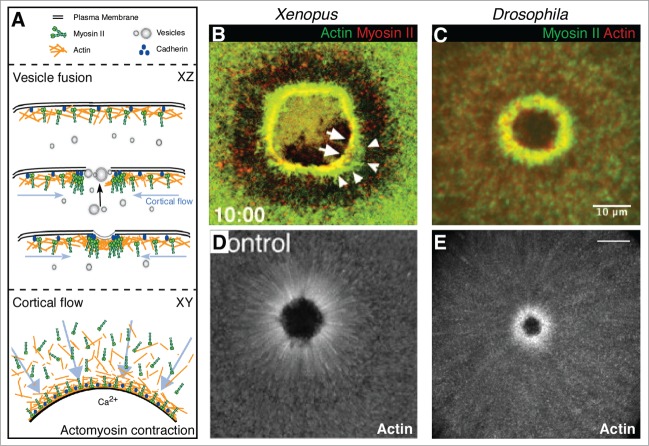
Figure 1.
Mechanisms of single cell repair. (A) Schematic depicting single cell repair mechanisms including vesicle fusion and cortical cytoskeleton remodeling by cortical flow and actomyosin contraction. While XY views depict the overall repair process, a 'behind the scenes' account of processes such as vesicles trafficking toward the wound from an internal membrane source are visible in XZ views (black arrow). Arrows (blue) in XY and XZ views show cortical flow as polarized trafficking of actin and myosin toward the wound. (B-D) Fluorescent micrographs of actomyosin contraction (B and C) or cortical flow (D and E) in single cell repair. An actomyosin array is formed at the wound edge in the Xenopus oocyte ((B) © 2001 Rockefeller University Press. Originally published in Journal of Cell Biology 154: 785–797) and the Drosophila syncytial embryo (C). Cortical flow of actin contributes to cortical remodeling in Xenopus (D; © 2005 Rockefeller University Press. Originally published in Journal of Cell Biology 168: 429–439) and Drosophila (E) single cell repair.

