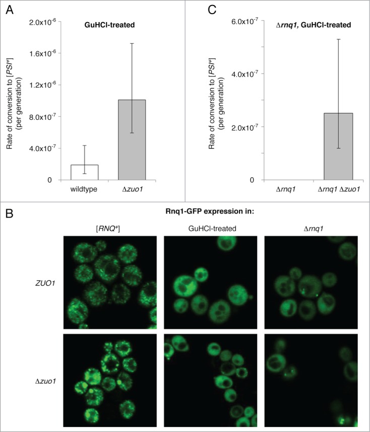
Figure 2.

Loss of RAC function bypasses the [PIN+] requirement in de novo [PSI+] formation. (A) Rates of spontaneous prion formation in wildtype and Δzuo1 strains that were cured to [rnq−] by treatment with GuHCl. Prion formation rates and 95% confidence intervals were calculated as for Figure 1B. (B) The aggregation state of Rnq1 was assessed in cells with (top panels) and without (bottom panels) RAC function in [RNQ+] cells (left), GuHCl−treated cells (center), and Δrnq1 cells (right) by expression of Rnq1-GFP from the inducible Cup1 promoter followed by confocal microscopy. Strains transformed with p316 Cup1pr-Rnq1-GFP were diluted into synthetic medium lacking uracil and supplemented with CuSO4 (25 µM) and incubated at 30°C for 90 minutes prior to imaging. (C) Rates of spontaneous prion formation in Δrnq1 and Δrnq1 Δzuo1 strains that were cured to [pin−] by treatment with GuHCl. Prion formation rates and 95% confidence intervals were calculated as for Figure 1B.
