
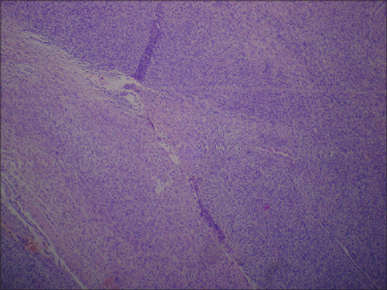
Figure 2.
Histopathology of the lesion in × 100 in H and E stain showing interlacing bundles of spindle cells with wavy nuclei. Spindle cells were intermixed with foci comprising dense bundles of collagen fibers giving a whirling pattern
Official websites use .gov
A
.gov website belongs to an official
government organization in the United States.
Secure .gov websites use HTTPS
A lock (
) or https:// means you've safely
connected to the .gov website. Share sensitive
information only on official, secure websites.
Histopathology of the lesion in × 100 in H and E stain showing interlacing bundles of spindle cells with wavy nuclei. Spindle cells were intermixed with foci comprising dense bundles of collagen fibers giving a whirling pattern