Figure 3.

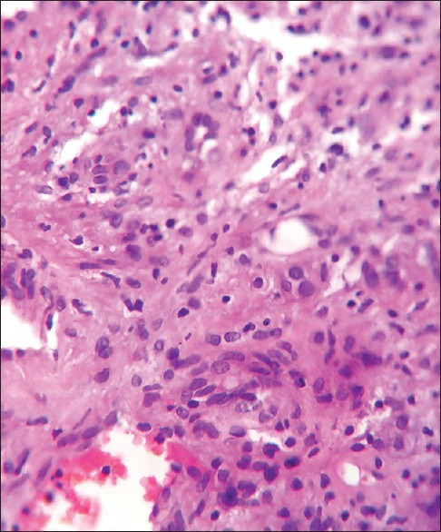
H and E staining showing multiple cavernous blood vessels alternating with cellular areas consisting of collapsed vascular spaces separated by spindle-shaped fibroblastic cells

H and E staining showing multiple cavernous blood vessels alternating with cellular areas consisting of collapsed vascular spaces separated by spindle-shaped fibroblastic cells