Figure 2.

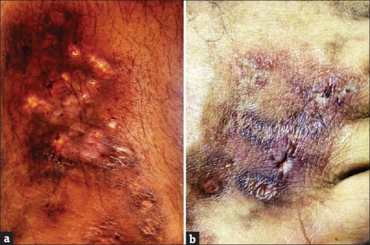
Close up images revealing (a) livedoid ulcers on leg long with atrophie blanche and (b) reticulate pattern of ulcers on dorsum of foot

Close up images revealing (a) livedoid ulcers on leg long with atrophie blanche and (b) reticulate pattern of ulcers on dorsum of foot