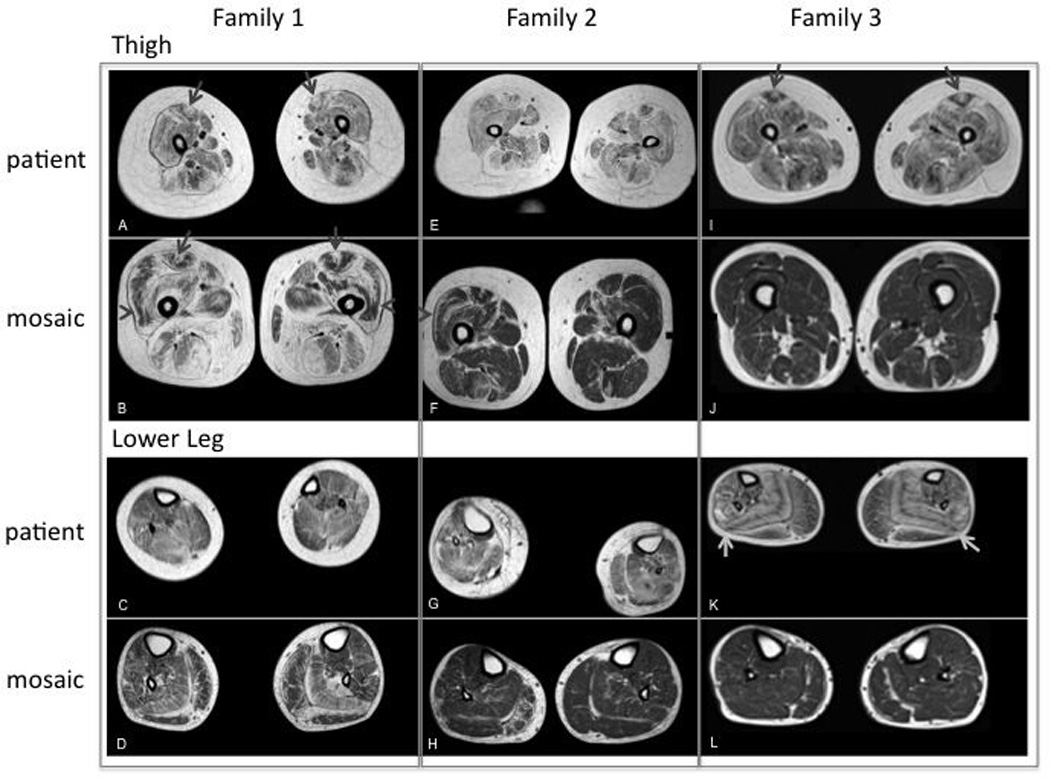
Figure 2.

Muscle MRI imaging for patient and mosaic parent from Family 1, 2 and 3. (A): Severe fibrofatty changes and atrophy in all muscles of the thigh. “Central shadow” is visible bilaterally (arrow); (B): Severe fibrofatty changes in the muscles of the posterior thigh (HAM) and adductor magnus. Moderate fibrofatty changes in the muscles of the anterior thigh with evidence of rimming (especially in RF and VL, arrow head) and "central shadow” bilaterally (arrow); (C): Mild fibrofatty changes in the muscles of the distal lower extremity; (D): Mild fibrofatty changes in most muscles of the distal lower extremity. More moderate fibrofatty changes are noted in the SOL and lateral gastrocnemius muscles bilaterally, which also exhibit atrophy; (E): Severe fibrofatty changes and atrophy of all muscles of the thigh. Adductor longus, hamstrings (HAM) and rectus femoris (RF) are most affected. No clear “central shadow” phenomenon; (F): Mild-moderate fibrofatty changes in the muscles of the thigh. On the right, there is a small central shadow in the RF. Rimming of fibrofatty changes in the RF and vastus lateralis (VL) are noted bilaterally; (G): Moderate fibrofatty changes and atrophy in all muscles of the distal lower extremity. (H): Mild fibrofatty changes in the soleus (SOL) bilaterally and peroneus (PER) and medial gastrocnemius (MG) muscle on the right; (I): Severe fibrofatty changes and moderate atrophy in all muscles of the thigh. “Central shadow” in the RF bilaterally; (J): mild fibrofatty changes in the HAM. No rimming or central cloud; (K): Moderate fibrofatty changes are severe in the SOL and PER muscle. Notes striped appearance of the SOL because of rimming with central preservation of muscle. Mild-moderate changes are noted in the remaining anterior and posterior muscles of the distal lower extremity; (L): Mild fibrofatty changes in the MG muscles bilaterally and PER on the right.
