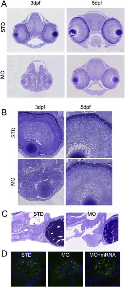
Fig. 4.
Knockdown of poc1b disrupts the morphogenesis of cilia in retina, heart and Kupffer's vesicle. (A) The whole head histology through coronal planes at 3 dpf and 5 dpf fish was illustrated. The head and brain size in poc1b morphants were approximately one-third of the size in control fish; and the size of eye was moderately smaller, in comparison to control. (B) The higher magnification views of the morphant retinas revealed no clear retina lamination at 3 dpf, but shortened and disorganized photoreceptor outer segments at 5 dpf in the retinas of poc1b morphants, with otherwise clear but thinner laminae. (C) In the control group, heart atrium and ventricle were developed normally with normal connection between them. However, in poc1b TB morphants, zebrafish larvae presented dilated ventricle, which have no proper connect with the atrium. (D) The Kupffer's vesicle cilia stained by acetylated alpha tubulin antibody in green were shortened and the morphology of the cilia was disrupted in poc1b TB morphants (middle panel). When co-inject poc1b MO with normal POC1B mRNA, the Kupffer's vesicle cilia disruption was partially rescued (right panel).

