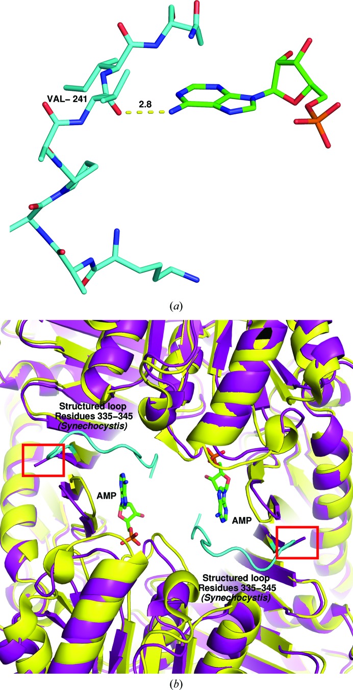
Figure 3.
The AMP-binding loop found in the Synechocystis sp. PCC 6803 structure is not visible in the T. elongatus electron density in the absence of AMP. (a) Stick model of Synechocystis sp. PCC 6803 residues 335–345 (cyan), which form a structured loop in the presence of AMP (green). The backbone carboxyl of Val341 forms a hydrogen bond to AMP. (b) AMP (green) is bound only in the Synechocystis sp. PCC 6803 structure (yellow cartoon). The final ten residues (structured loop) of the Synechocystis sp. PCC 6803 structure are shown in cyan as in (a) to indicate that they form part of the AMP-binding region. The purple cartoon is the T. elongatus FBP/SBPase. The red box indicates the end of the T. elongatus structure, as the final residues are not visible in the electron density.

