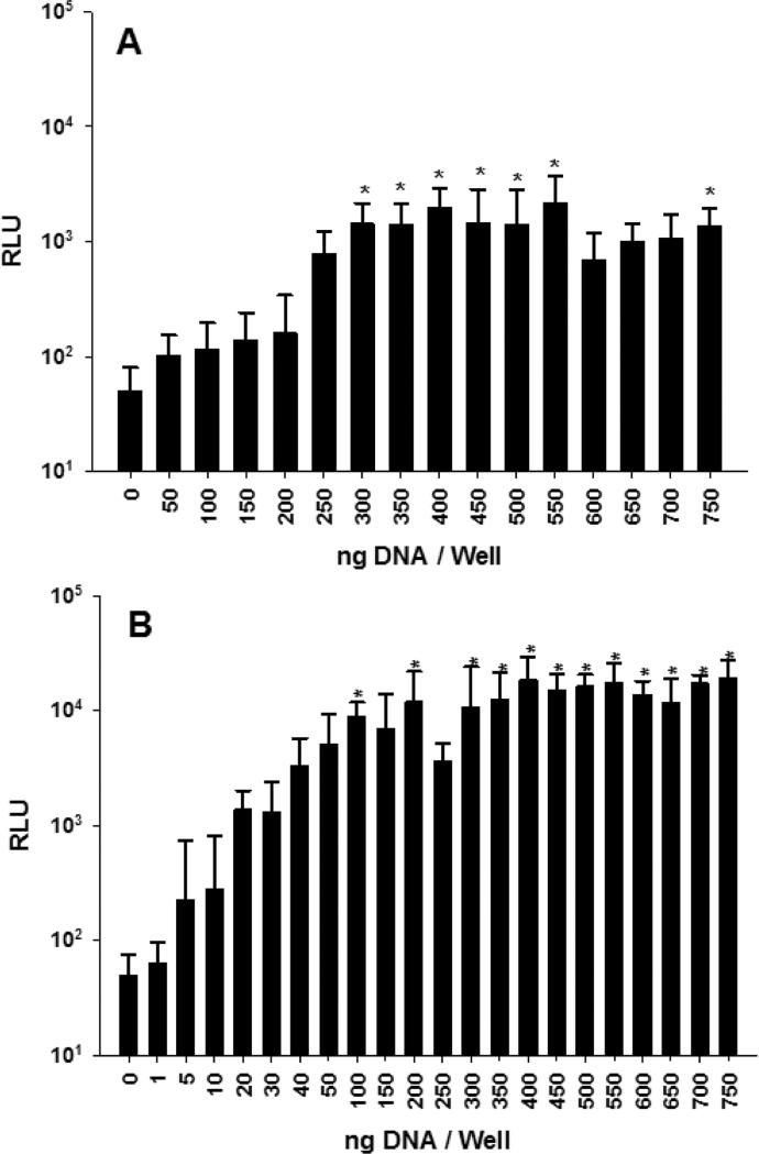
Figure 8.
PEI and CaPO4 Transfection of Primary Hepatocytes in 384-Well Plates. Panel A illustrates the transfection of 250 primary hepatocytes (45 μL) per well with 0-750 ng of PEI gWiz-Luc (N:P 7). At 24 hr 10 μL of ONE-Glo was added and bioluminescence was measured at 5 min (n = 9). Panel B illustrates the transfection of primary hepatocytes (45 μL) plated in 384-well plates at 250 cells per well with 250 ng of CaPO4 calcium phosphate. At 24 hr 10 μL of ONE-Glo was added and bioluminescence was measured at 5 min (n = 9). * indicates p ≤ 0.05 relative to 0 DNA dose.

