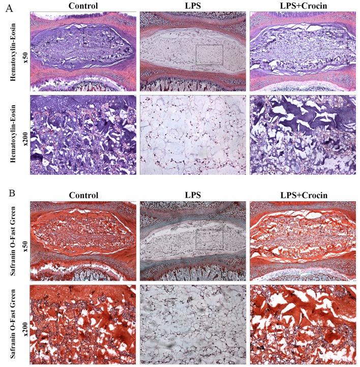
Figure 5.
Histological analysis of intervertebral discs (IVDs) cultured ex vivo for the assessment of IVD degeneration. (A) Hematoxylin-eosin (H&E)-stained images under low (×50) and high (×200) magnification. (B) Safranin O-fast green-stained images under low (×50) and high (×200) magnification. LPS, lipopolysaccharide. The high magnfication images are representative of the insets in the low magnification images.

