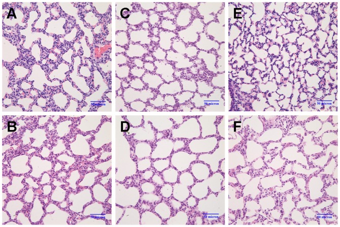
Figure 1.
Changes in lung morphology. Lung sections were stained with H&E and images were captured under a light microscope (magnification, ×40). (A andB) On postnatal day 3, the alveolar walls were slightly thicker in the model group (M3d) compared to the control group (C3d). (C and D) On postnatal day 7, the number of alveoli had markedly decreased, secondary septa formation was reduced, and the alveolar size was slightly increased in the model group (M7d) compared to the control group (C7d). (E and F) On postnatal day 14, the following changes between the 2 groups became more apparent: thickening of the alveolar wall, decrease in the number of alveoli, reduced numbers of secondary septa, and alveolar structural abnormalities (control group, C14d; model group, M14d). Model group refers to the bronchopulmonary dysplasia (BPD) group.

