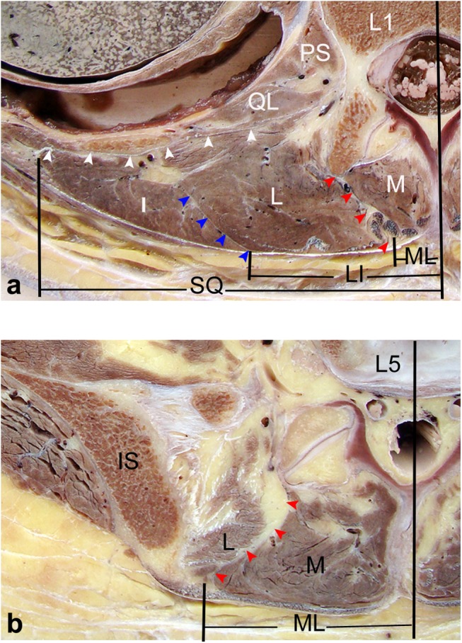
Fig 5. Horizontal views of the posterior lumbar spine in anatomical sections obtained from cadavers.

(a) Through the upper level of the lumbar spine (L1), the muscles in the posterior lumbar region were divided into three parts: the medial part (multifidus), middle part (longissimus) and lateral part (iliocostalis). The longissimus and iliocostalis were the two parts of the sacrospinalis. The red arrows point to the ML, which is a natural corridor filled with adipose tissue between the multifidus and longissimus. The blue arrows show the LI, which is a thin fascia between the longissimus and iliocostalis. The white arrows point to SQ between the sacrospinalis and quadratus lumborum. (b) Through the lower level of the lumbar spine (L5), the ML was well defined, and slightly more lateral to the midline. LI and SQ could not be identified. M: multifidus; L: longissimus; I: iliocostalis; IS: iliac spine; QL: quadratus lumborum; PS: psoas major.
