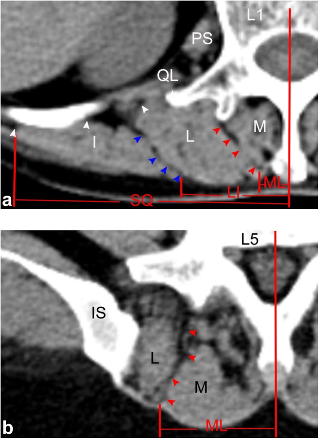
Fig 6. Horizontal views of the posterior lumbar spine in CT images.

(a) Through the upper level of the lumbar spine (L1), ML (red arrows), SQ (white arrows) and LI (blue arrows) were identified by line-shaped low densities within the paraspinal muscles. (b) Through the lower level of the lumbar spine (L5), only the ML was identified. M: multifidus; L: longissimus; I: iliocostalis; IS: iliac spine; QL: quadratus lumborum; PS: psoas major.
