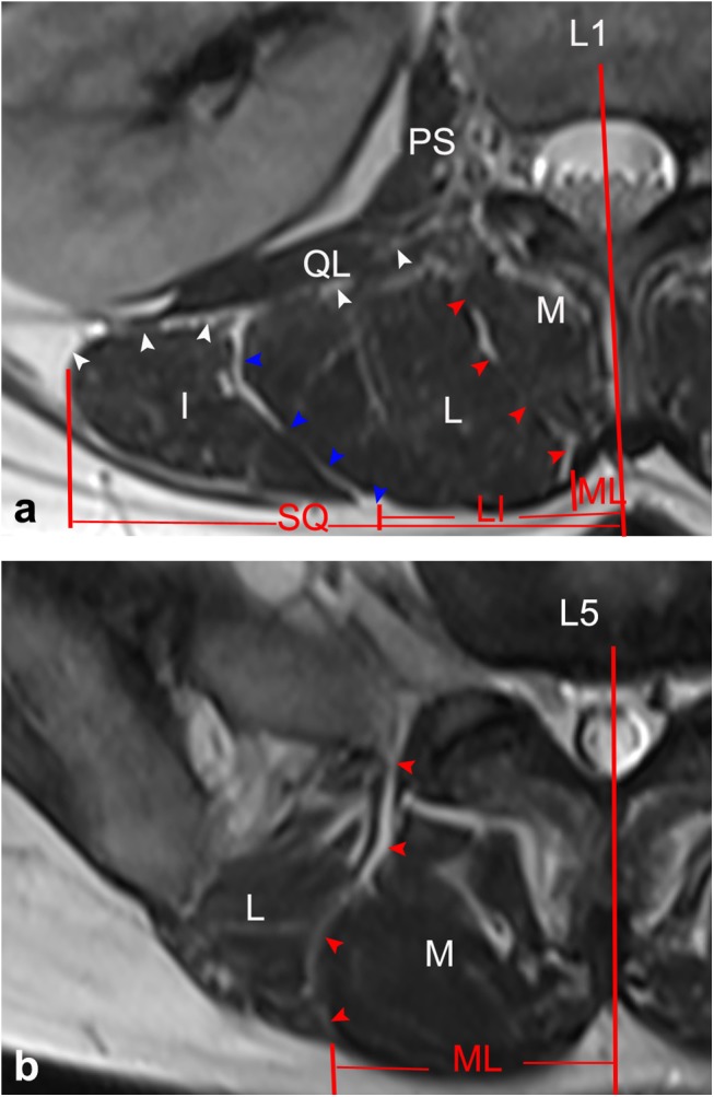
Fig 7. Horizontal views of the posterior lumbar spine in MRI images.

(a) Through the upper level of the lumbar spine (L1), ML (red arrows), SQ (white arrows) and LI (blue arrows) were identified by lines of high signal intensity inserted into the paraspinal muscles. (b) Through the lower level of the lumbar spine (L5), only the ML was identified. M: multifidus; L: longissimus; I: iliocostalis; IS: iliac spine; QL: quadratus lumborum; PS: psoas major.
