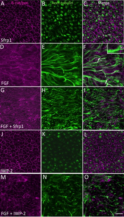
Figure 3.

Sfrp1 or IWP-2 inhibits FGF-induced fiber elongation. Explants were cultured in 4 μg/mL Sfrp1 (A–C), 200 ng/mL FGF-2 (D–F), in the presence of both FGF and Sfrp1 (G–I), or in the presence of both FGF and IWP-2 (20 μM, J–L) for 4 days. β-catenin and pericentrin immunoreactivity (A, D, G, J) localizes cell margins and centrosomes, respectively. Acetylated-tubulin reactivity (B, E, H, K) localizes stabilized microtubules and helps visualize extent of cell elongation. In Sfrp1 alone, the cells maintain the cobblestone-like arrangement characteristic of lens epithelial cells in vivo (A–C). In FGF alone, cells show extensive elongation (D–F). Pericentrin reactivity localizes the centrosome and identifies the apical tip of each cell (F, inset) and groups of elongated cells are often seen to point in a similar direction (F, arrows). When Sfrp1 (G–I) or IWP-2 (M–O) is included with FGF, elongation is not as extensive as in explants with FGF alone. Scale bar: 20 μm.
