Figure 5.

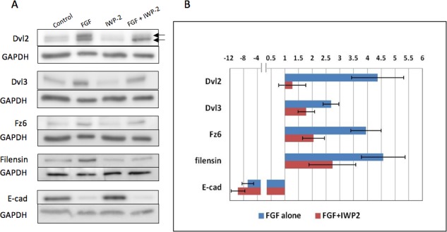
IWP-2 inhibits FGF-induced fiber differentiation. (A) Western blots for Dvl2, Dvl3, Fz6, Filensin, and E-cadherin in explants after 4 days exposure to either 200 ng/mL FGF-2, IWP-2 alone, FGF-2/IWP-2, or no FGF with DMSO vehicle only (controls). Dvl2, Dvl3, Fz6, and filensin are all upregulated after 4 days exposure to FGF. In contrast, E-cadherin expression is reduced. Explants cultured with IWP-2 show similar band densities for E-cadherin as in controls (no FGF with DMSO vehicle only). When IWP-2 is included with FGF, band densities for Dvl2, Dvl3, Fz6, and filensin are all reduced in comparison with explants cultured in FGF alone. E-cadherin is relatively unaffected by the FGF/IWP-2 combination. Dvl2 is present in two bands (arrows); the upper band being the phosphorylated form of Dvl2. In explants cultured with FGF/IWP-2, the upper band in particular is reduced in comparison to explants cultured in FGF alone. (B) The histogram shows band density measurements for Dvl2 (upper phosphorylated band only), Dvl3, Fz6, filensin, and E-cadherin. In all cases, GAPDH loading controls were used to normalize the levels of protein detected and the results were presented as fold changes relative to the respective controls. Data presented shows the mean ± SEM (n = 4). Similar to the results shown in Figure 1, FGF induced about a 2- to 4-fold increase in expression of Dvl2 (phosphorylated), Dvl3, Fz6, and filensin. Also, as before, E-cadherin was reduced in FGF-treated explants. In all cases, except for E-cadherin, the presence of IWP-2 significantly reduced protein expression when compared to groups treated with FGF alone (P = ≤ 0.05, ANOVA with Tukey's test). Notably, Dvl2 showed a significant decrease in the density of the upper phosphorylated (active Dvl2) band indicating reduced Dvl2 signaling activity.
