
Fig. 8.
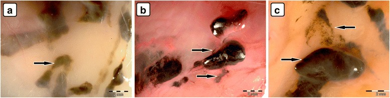
Visualization of CNP aggregates under a stereomicroscope after multiple intraperitoneal injections. Diamond (a), graphite (b), and graphene oxide (c) nanoparticles accumulated in a similar way; large aggregates up to 10 mm and small dots around 1 μm were formed. Black arrows indicate CNP aggregates