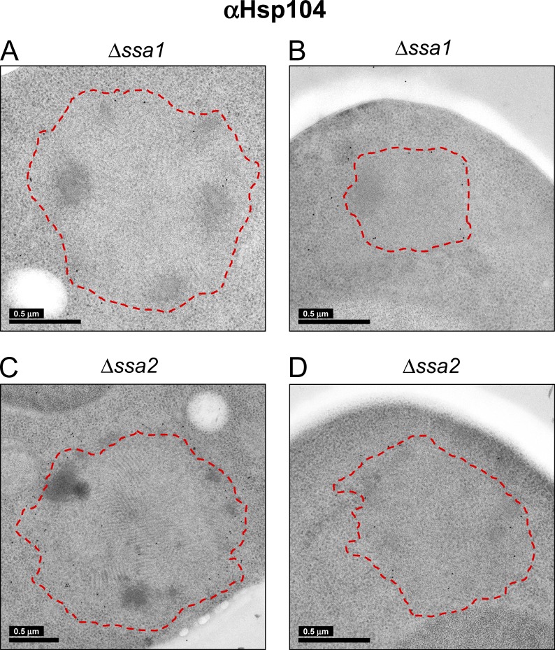
Figure 7.
Immunogold detection of Hsp104 on NM-YFP assemblies in Hsp70 knockout cell sections. (A–D) Representative EM projection images of immunogold-labeled cell sections of Δssa1 (A and B) and Δssa2 (C and D) cells, as indicated. Detection was with an α-Hsp104 antibody. The red outlines, encompassing entire NM-YFP dot aggregates, were manually traced at the interface between the peripheral unstructured zone of the aggregates and the ribosomes in the cytosol.

