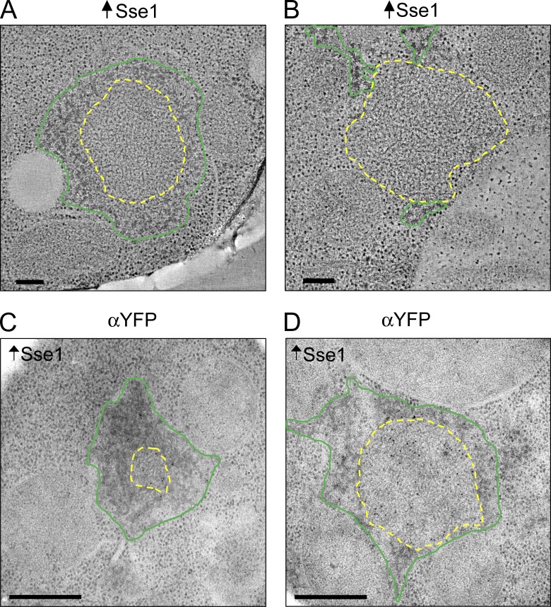
Figure 9.
Sse1 overexpression disrupts NM-YFP aggregate organization. (A and B) Representative tomographic slices from reconstructions of NM-YFP dots in cell sections of HM20-embedded cells with a wild-type chaperone complement and overexpressing Sse1 from plasmid pSse1. Normal fibrillar regions are outlined in yellow, manually traced at the interface between the fibrils and the surrounding cytosolic ribosomes and/or mesh-like amorphous aggregates. The mesh-like amorphous material was manually outlined in green. Bars, 200 nm. (C and D) Representative EM projection images of immunogold-labeled HM20-embedded cell sections of cells with a wild-type chaperone complement and overexpressing Sse1. Sections were labeled with an α-YFP antibody to detect NM-YFP. The mesh-like amorphous material was sparsely labeled, suggesting that it contained mostly other aggregates. Fibrils are outlined in yellow and the amorphous material is outline in green, as described for A and B. Bars, 0.5 µm.

