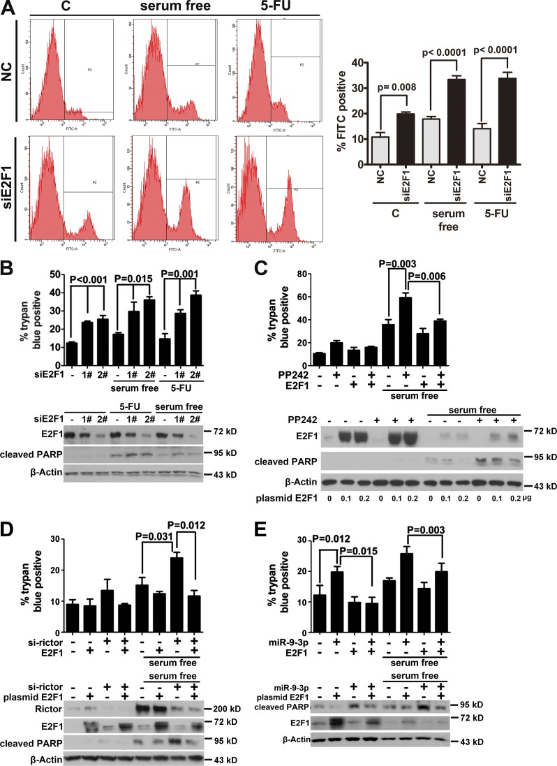
Figure 4.
miR-9-3p acts downstream of mTORC2 and triggers apoptosis by targeting E2F1. (A) MCF-7 cells were transfected with a pool of two siRNAs (1:1) targeting different regions of E2F1 mRNA. After 36 h, cells were serum starved or treated with 400-µM 5-FU for an additional 24 h, harvested, and labeled with Annexin V–FITC and propidium iodide for analysis of apoptosis. The data shown are from a single representative experiment out of three repeats. (B) MCF-7 cells were transfected separately with the two siRNAs and treated as in A, followed by trypan blue staining (top) or Western blotting (bottom). (C) MCF-7 cells were transfected with vehicle or WT E2F1 in the absence or presence of PP242, as indicated. 12 h after transfection, cells were serum starved for an additional 24 h and harvested for either trypan blue staining (top) or Western blotting (bottom). (D) MCF-7 cells were sequentially transfected with Rictor siRNA and WT E2F1. After 24 h of serum starvation, the effects of ectopic E2F1 expression on Rictor knockdown were monitored via trypan blue staining (top) and Western blotting (bottom). (E) MCF-7 cells were sequentially transfected with miR-9-3p and E2F1, followed by assay as described in C. Error bars represent mean values ± SEM. C, control; NC, negative control.

