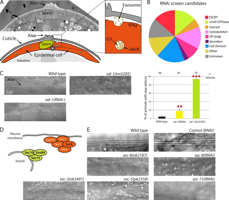
Figure 1.
RAL-1 GTPase or exocyst deficiency induces alae defects. (A) C. elegans epidermal cells contain MVBs, which can fuse with the apical plasma membrane and liberate exosomes. These exosomes are integrated in the cuticle and contribute to the formation of the alae. (B) An RNAi-based screen identified 73 genes required for alae formation. (C) Disruption of ral-1 by RNAi, or by the null allele ral-1(tm5205), leads to alae defects. The number of animals is shown at the top of the graph. **, P < 0.05; ***, P < 0.01. (D) Schematic representation of the exocyst complex involved in plasma membrane attachment of secretory vesicles. Subunits found in the screen are in black. (E) Alae defects observed after disruption of several members of the exocyst complex in mutants or by RNAi.

