Figure 2.
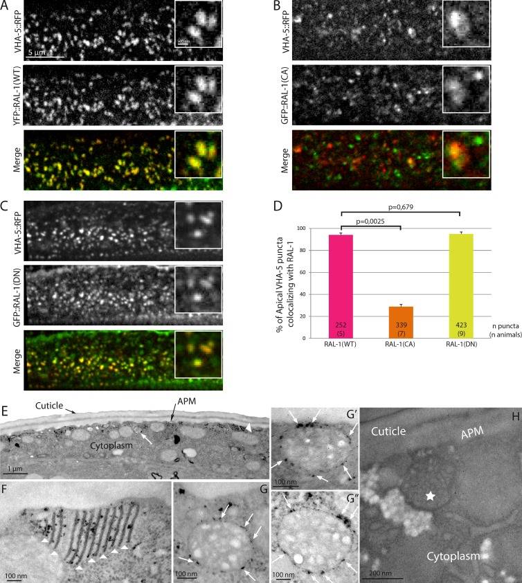
RAL-1 localizes at the surface of MVBs. (A–D) WT (RAL-1(WT); A) and dominant negative (RAL-1(DN); C) versions of RAL-1, but not the constitutively active version (RAL-1(CA); B), colocalize fully with VHA-5 in the epidermis at the time of alae formation, as shown in the quantification (D). In D, the numbers inside the bars indicate the number of puncta (number of animals). (E–H) APEX::RAL-1(DN) shows DAB staining both at apical membrane stacks (E and F, arrowheads) and at the external surface of MVBs (E and G–G’’, arrows). Animals expressing no APEX tag treated with DAB show no staining (H). The star indicates a MVB. APM, apical plasma membrane.

