Fig. 3.
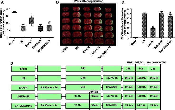
Neurological scores and infarct volumes at 72 h after reperfusion in the rats. a Garcia scores were tested at 72 h after reperfusion. b Representative brain infarct size indicated by TTC staining at 72 h after reperfusion. c Statistical analysis of the infarct size in every group (% of contralateral hemisphere) (*p < 0.05 vs. I/R; #p < 0.05 vs. EA + I/R). d Experimental protocol, SD rats were randomly divided into five groups (n = 8 each): Sham, I/R, 2ME2 + I/R, EA + I/R, and EA + 2ME2 + I/R group. At 30 min before MCAO, the 2ME2 were administered. At 72 h after ischemia/reperfusion, neurological function scores, and infarct volumes were assessed in every group. At 24 h after ischemia/reperfusion, TUNEL staining, the expression of Bcl2 and Bax was tested to evaluate neuronal apoptosis in every group

