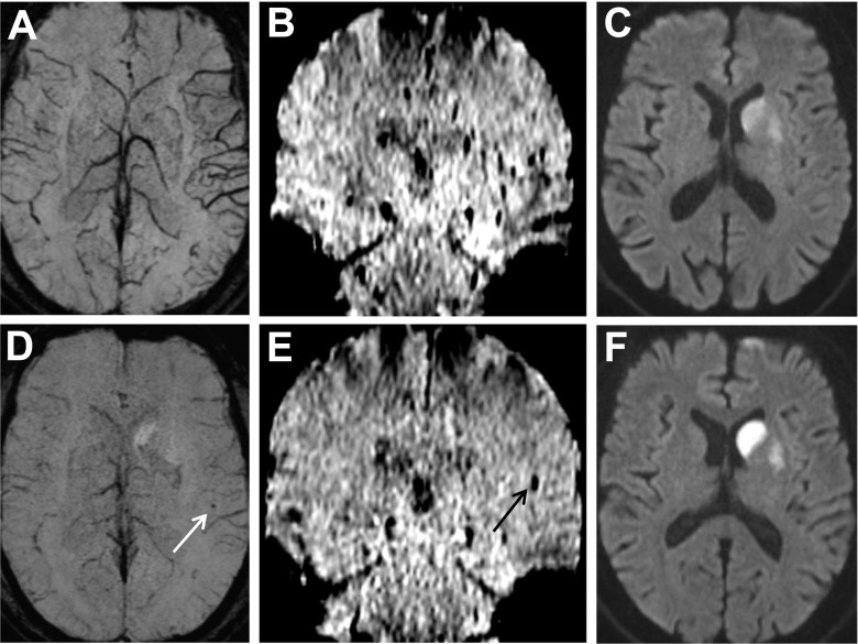
Fig. 1.
A 72-year-old man with mild aphasia and right-sided hemiparesis (NIHSS score 5). MRI shows left proximal MCA occlusion. On initial SWI, prominent veins in the region of the left Sylvian fissure are seen (a [axial, mIP] and b [coronal]). DWI shows diffusion restriction in the basal ganglia (c). Post-interventional SWI demonstrates a punctate signal drop corresponding to a peri-interventional embolus (d [white arrow, mIP] and e [black arrow]). The embolus is located outside of the infarcted area as seen on post-interventional DWI (f)

