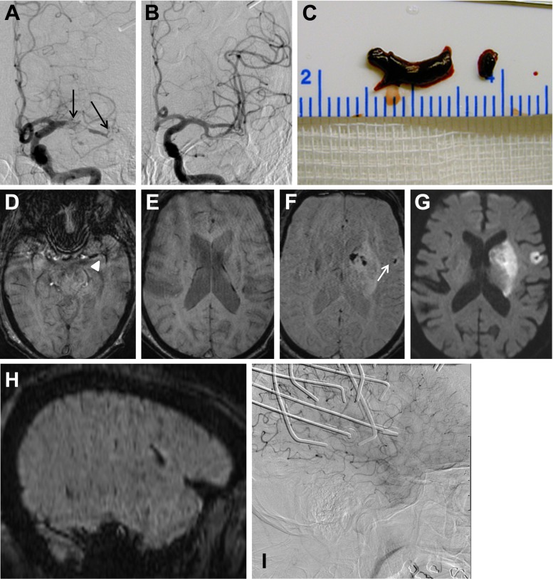
Fig. 2.
A 83-year-old man with right-sided hemiparesis (NIHSS score 21). Two separate thrombi in the left proximal M1 segment and the MCA bifurcation are seen on DSA (a, black arrows). Pre-interventional SWI shows thrombotic material in the proximal M1 segment and MCA bifurcation extending in an M2 branch (measuring 10 and 9 mm on SWI; d, white arrowhead) but no additional distal fragments (e). Two thrombus fragments were retrieved (c). Final biplane DSA control was rated as TICI 3 reperfusion (b). Post-interventional SWI detected one EE (f, white arrow) with an associated ischemic lesion on DWI (g). The sagittal reformatted projection of the SWI confirms the extracerebral location of the EE and its orientation along a suprasylvian MCA branch (h). Lateral DSA shows stagnating flow in the corresponding MCA branch with discretely diminished parenchymal blush during this phase of the DSA (i). (Artifacts are produced by simultaneous recording of NIRS)

