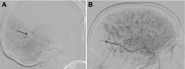
Fig. 4.
The DSA of the dominant vertebral artery in this 79-year-old woman with occlusion of the internal carotid artery shows the retrograde filling of the posterior parietal branches of the MCA (a). Due to the still active collaterals from the vertebrobasilar system, not visible in the selective DSA, the interventional team rated this as TICI 2b based on the stagnating flow and diminished temporooccipital blush (b). Quality control MR the next day found neither EE nor DWI lesions in this area

