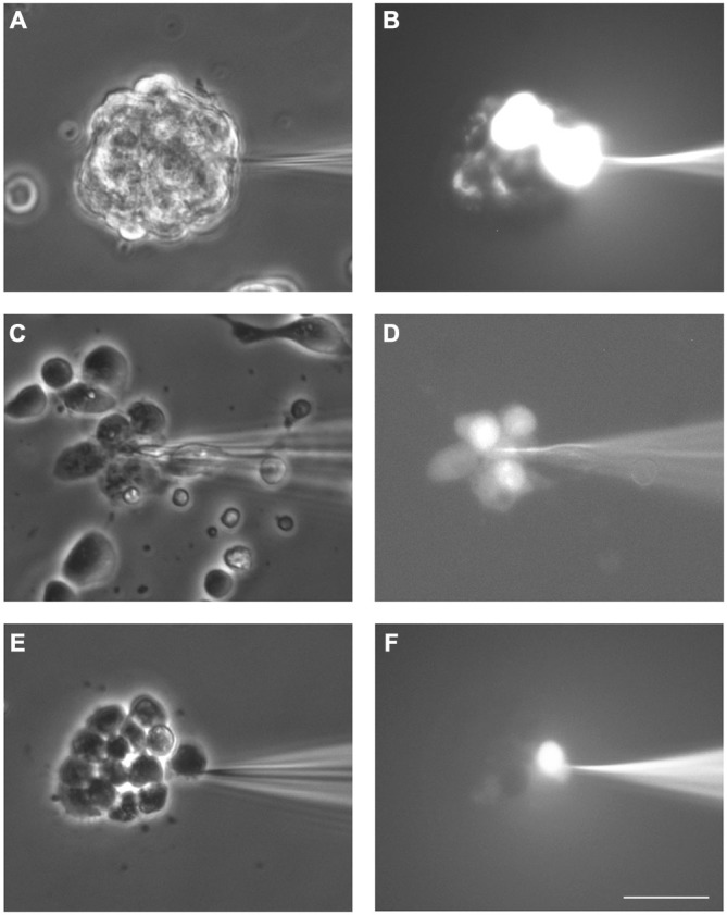
Figure 2.

Dye coupling between cells in SVZ neurospheres. Examples of dye coupling 3 min after Lucifer yellow (LY) microinjection into a cell of a non-disaggregated neurosphere (A, B) or into a neurosphere-derived cell (C, D). After addition of the gap junction blocker octanol (750 μM), microinjected LY did not spread to neighboring neurosphere-derived cells (E, F). (A,C) and (E) show the corresponding phase-contrast views of the fields of the representative pictures of LY microinjections shown in (B,D) and (F), respectively. Bar: 50 μm.
