Figure 4.
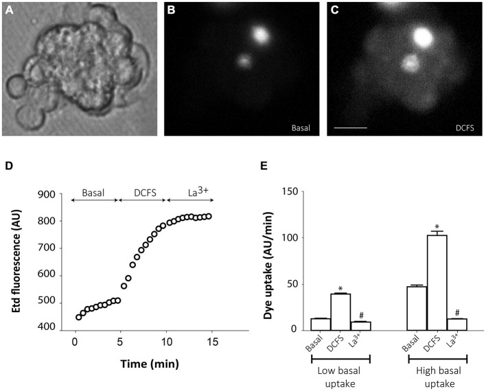
Connexin hemichannel activity in SVZ neurosphere-derived cells. Representative fluorescence images of a small cluster of neurosphere-derived cells incubated in a solution containing 5 μM ethidium (Etd) under basal conditions (B) and after bathing with divalent cation-free solution (DCFS) to increase the open probability of connexin hemichannels (C). (A) shows the corresponding phase-contrast image. Brightest cells in B are cells with high basal Etd uptake in contrast to the majority of neurosphere-derived cells that presented very low basal Etd uptake. Bar: 25 μm. (D): Time-lapse Etd uptake (in arbitrary units, AU) under basal conditions (Basal; first 5 min), after incubation in DCFS (DCFS, following 5 min) and after addition of the connexin hemichannel blocker 200 μM La3+ (La3+, following 5 min). Each point corresponds to the mean of 10 cells with low basal Etd uptake from one experiment. (E): Etd uptake rates (in AU/min) in neurosphere-derived cells measured under basal conditions (Basal), after incubation in DCFS (DCFS) and after La3+ addition (La3+) in low basal Etd uptake cells and in high basal Etd uptake cells. Data are presented as mean ± SEM from three independent experiments. *p < 0.05 compared to basal conditions. #p < 0.05 compared to DCFS condition (one-way ANOVA followed by Tukey’s post hoc test).

