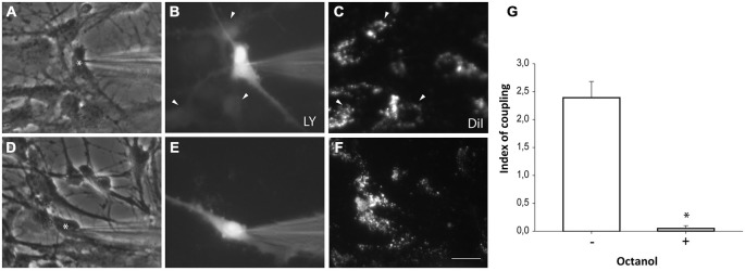
Figure 5.

Neurosphere-derived cells and astrocytes establish gap junctional communication. Photomicrographs showing examples of LY microinjections in co-cultures of neurosphere-derived cells and primary astrocytes. Astrocytes were labeled with the lipophilic dye DiI before the co-culture to allow their identification. LY was microinjected in neurosphere-derived cells and the transfer to adjacent DiI-labeled cells was evaluated three minutes later. (A): Phase-contrast view of the fields shown in (B,C). The asterisk denotes the cell microinjected with LY. (B): LY microinjection in a neurosphere-derived cell and dye transfer to adjacent cells (arrowheads). (C): DiI-labeled cells of the same field shown in (B). Cells that received the dye are indicated with arrowheads. (D): Phase-contrast view of the fields shown in (E,F). The asterisk denotes the cell microinjected with LY. (E): LY microinjection in a neurosphere-derived cell after addition of the gap junction blocker octanol (750 μM). A lack of dye transfer to adjacent cells is observed. (F): DiI-labeled cells of the same field shown in (E). Bar: 50 μm. (G). Index of coupling, calculated as the mean number of cells that received LY in every injection, in co-cultures of neurosphere-derived cells and astrocytes in control conditions (−) and after addition of octanol to the extracellular saline solution (+). Results are the mean ± SEM of 96 cells from nine independent experiments). *p < 0.05 compared to control conditions (Student’s t test).
