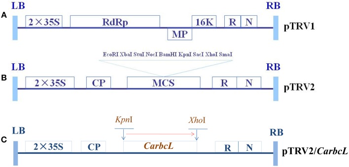
Figure 1.
Schematic representation of TRV and recombinant TRV vectors carrying the target genes. (A) The structure of pTRV1; (B) The structure of pTRV2; (C) The structure of TRV-CarbcL. LB, left border of the T-DNA; RB, right border of the T-DNA; 2 × 35S: two copies of the cauliflower mosaic virus 35S promoter; CP, coat protein; RdRp, RNA-dependent RNA polymerase; MP, movement protein; 16K, 16 KDa protein; R, ribozyme; N, NOS terminator; and MCS, multiple cloning site.

