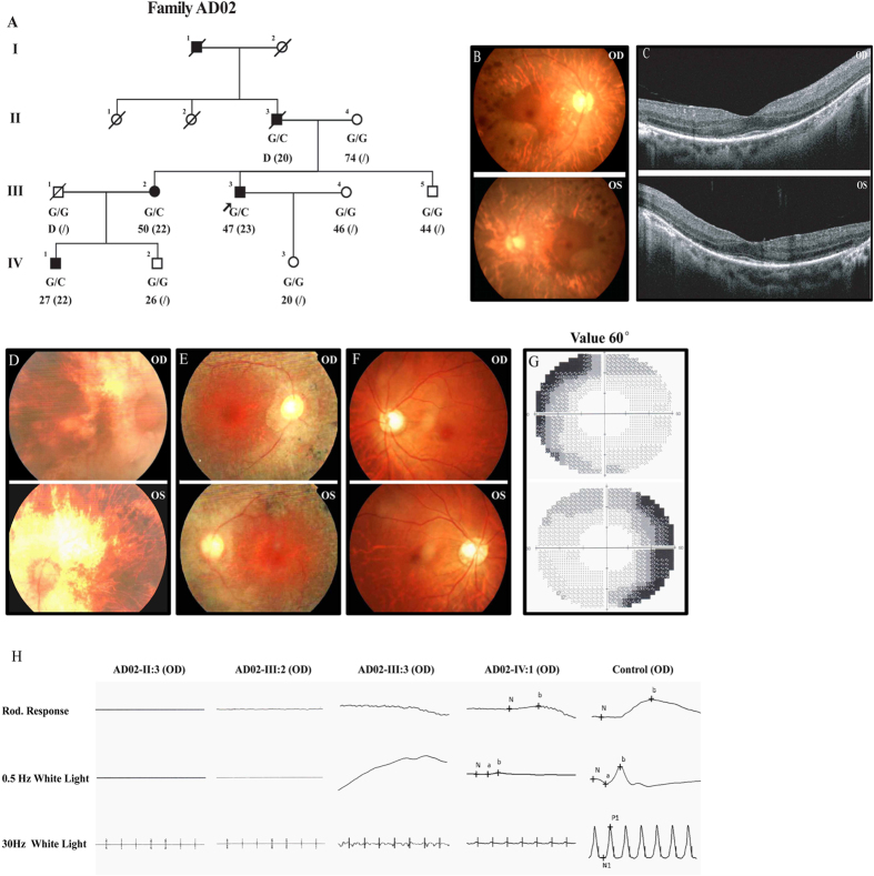
Figure 1. Detailed clinical evaluations of patients from family AD02.
(A) The pedigree of family AD02 indicates a dominant inheritance pattern of four generations. Genotypes, current and onset ages of RP (inside parentheses) are shown below the pedigree symbols. (B,D–F) Fundus photos demonstrate typical RP changes including waxy optic disks, artery attenuations, pigment deposits, and macular degeneration, in both eyes of patient AD02-III:3 (B), AD02-II:3 (D) and patient AD02-III:2 (E), while the fundus of patient AD02-IV:1 was normal (F). (C) Macular degeneration is also indicated by OCT examinations, which reveal attenuated ONL and RPE with complete loss of OS and IS. (G) Peripheral vision loss is revealed by automated visual field examination of patient AD02-IV:1. (H) Scotopic and photopic ERG responses of patients AD02-II:3, III:2, and III:3 are undetectable, while are significantly reduced for patient AD02-IV:1. ERG responses of a negative control are also presented.

