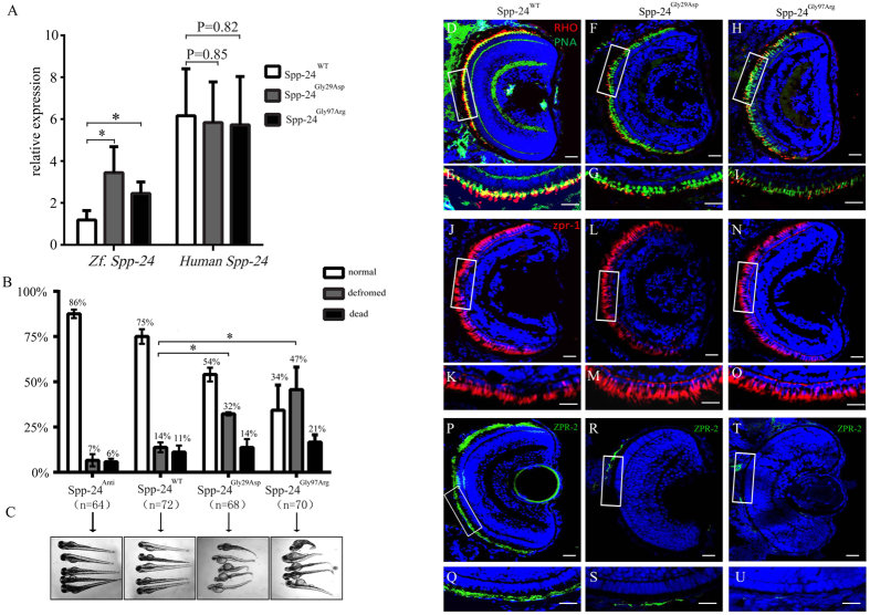
Figure 4. Deleterious effects identified in zebrafish overexpressing Spp-24 mutants.
(A) Expressions of endogenous spp2 (Zf.) and exogenous SPP2 (Human) in zebrafish at 2 days post fertilization (dpf) after injection of Spp-24WT, Spp-24Gly29Asp and Spp-24Gly97Arg were determined by Q-PCR and were relative to the expression of spp2 in uninjected larvae. (Spp-24Gly29Asp VS Spp-24WT, P = 0.0463; Spp-24Gly97Arg VS Spp-24WT, P = 0.0379). (B) Quantification of normal, deformed, and dead zebrafish injected with different mRNAs from 2 to 4 dpf. n: total numbers of injected zebrafish from triple experiments. (Spp-24Gly29Asp VS Spp-24WT, P = 0.0142; Spp-24Gly97Arg VS Spp-24WT, P = 0.0105). (C) Morphological changes in zebrafish injected with different mRNAs at 4 dpf. Most zebrafish injected with Spp-24Anti and Spp-24WT is relatively normal, while significant systemic deformations are revealed in groups injected with Spp-24Gly29Asp and Spp-24Gly97Arg. (D–U) Immunostaining of rhodopsin (D–I), peanut agglutinin (PNA) lectin (D–I), ZPR-1 (J–O), and ZPR-2 (P–U) on retinal frozen sections of zebrafish at 4 dpf from the four groups, including groups injected with Spp-24WT, Spp-24Gly29Asp and Spp-24Gly97Arg. Robust staining of rhodopsin is found in the rod IS/OS and cone OS layers in Spp-24WT -injected fish, respectively (D–E). Reactivity of rhodopsin (F–I) and ZPR-2 (Q–U) are decreased in Spp-24Gly29Asp and Spp-24Gly97Arg-injected zebrafish, while reactivities of ZPR-1 and PNA was clearly detected in the IS/OS layer of all zebrafish studied (D–O). The boxed areas in (D,F,H,J,L,N,P,R,T) were shown in higher magnification in (E,G,I,K,M,O,Q,S,U). Scale bar: 20 μm.

