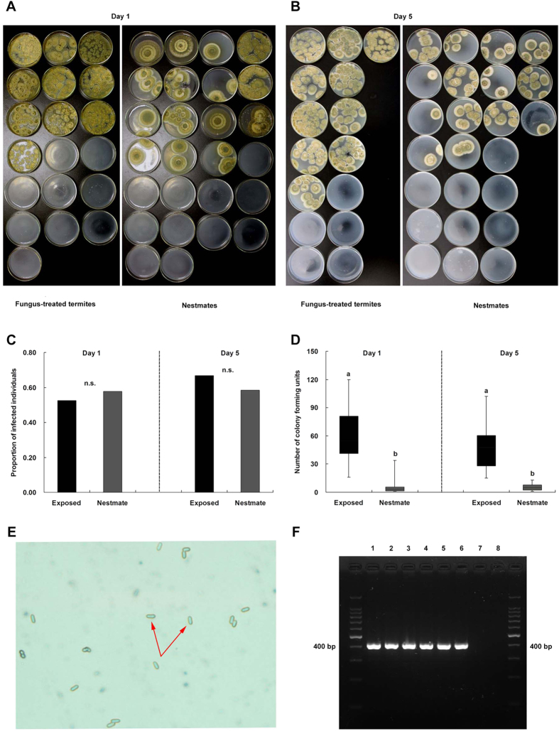
Figure 3. Nestmates experienced low-level M. anisopliae infections.
(A,B) Growth of CFUs from fungus-treated termites and their nestmates after 1 d and 5 d of social contact. (C,D) Infection levels of fungus-treated termites (black bars) and their nestmates (grey bars) including the proportion of fungal growth and the number of CFUs after 1 d and 5 d of social contact (Mann-Whitney U-test, p < 0.05). (E) Identification of CFUs from conidia of nestmates of fungus-treated termites as M. anisopliae by morphological determination. (F) Identification of CFUs as M. anisopliae by PCR using primers specific for M. anisopliae, including PCR products of DNA from M. anisopliae (lanes 1 and 2, positive controls), from CFUs of fungus-treated termites (lanes 3 and 4), from CFUs of nestmates of fungus-treated termites (lanes 5 and 6) and from Beauveria bassiana (lanes 7 and 8, negative controls).

