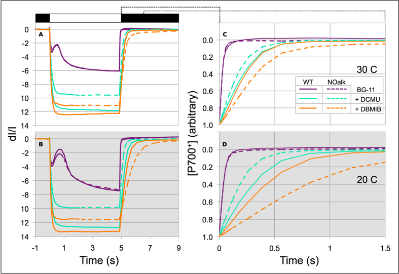
Figure 4. P700 redox kinetics for WT and noALK strains at 20 and 30 C.
Using a JTS-10 spectrophotometer, cell suspensions were dark-adapted and then exposed to a pulse of orange actinic light (to excite both PSII and PSI) for 5 seconds (white bar above panel (A)). The actinic light was then turned off (black bar above panel (A)). During this time-course, measuring flashes of 705 nm light probed the redox state of the P700 reaction center of PSI. Data were collected from cells that had been grown at 30 C, then measured at 30 C (panel (A,C)) or shifted to 20 C before measurement (panel (B,D)). Panels C and D show the details of re-reduction of P700+ in the dark for the experiments in panels A and B, respectively. Inhibitors of linear electron flow (10 μM DCMU, which inhibits transfer from PSII to the quinone pool), and linear + cyclic electron flow (1 μM DBMIB, which blocks cyt b6f) were added as indicated. Each trace is an average of 3 independent experiments.

