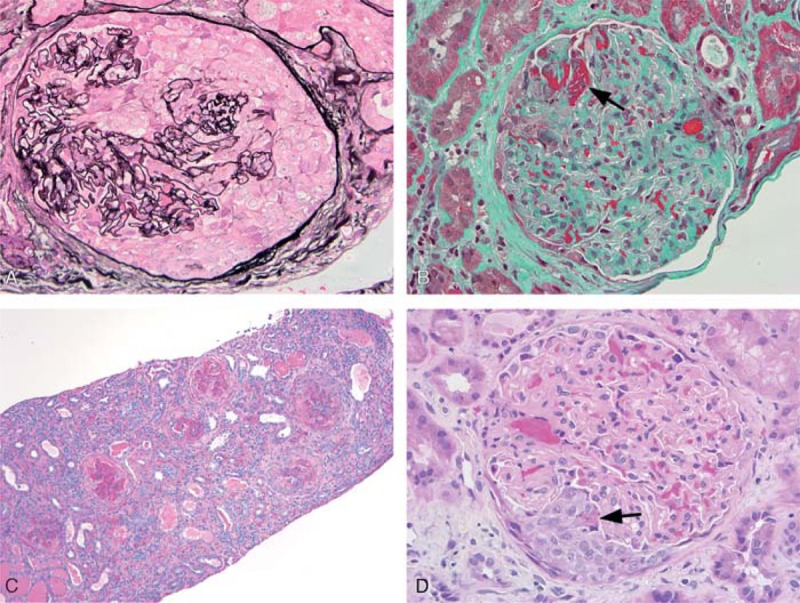
FIGURE 1.

Kidney biopsy histologic findings. A) Glomerulus from Case 1 with a large cellular crescent (methenamine silver-periodic acid-Schiff; 400x). B) Glomerulus from Case 1 showing fibrin (arrow) of a necrotizing lesion (trichrome; 400x). C) Medium power view of biopsy from Case 4 demonstrating a range of cellular to fibrocellular crescents. Features of mild interstitial fibrosis, tubular atrophy, and interstitial inflammation are also noted (periodic acid-Schiff; 100x). D) Glomerulus from Case 4 showing a small cellular crescent with associated fibrin of a necrotizing lesion (arrow) (hematoxylin & eosin; 400x).
