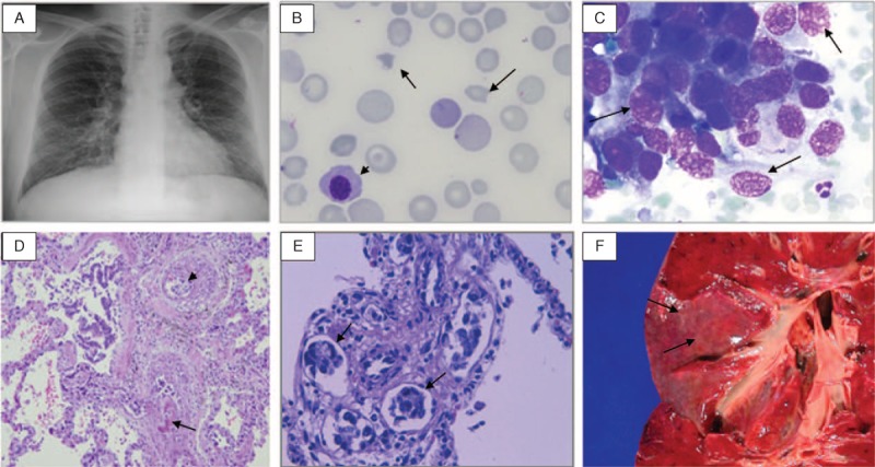
FIGURE 1.

A. Chest X-ray with interstitial bilateral infiltrates (Case 1). B. Schistocytes (arrows) and an erythroblast (arrowhead) in the peripheral blood smear (Case 3). C. Neoplastic cells in bone marrow (arrows) (Case 3). D. Blood vessels with eccentric intimal fibrosis (arrow), intravascular fibrin thrombi, and recanalization and intraluminal emboli of neoplastic cells (arrowhead) (hematoxylin and eosin, original magnification × 100) (Case 3). E. Carcinoma emboli in perivascular lymphatic vessels (arrows) (hematoxylin and eosin, × 400) (Case 1). F. Autopsy specimen of the lung showing prominent lymphatic vessels (carcinomatous lymphangitis) in pleural surface as fine white lines (Case 3).
