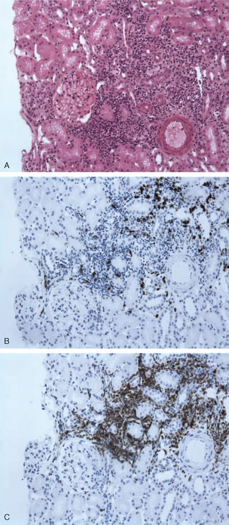
FIGURE 1.

Kidney biopsy showing nontumoral interstitial infiltration from a patient with MCNS and marginal zone lymphoma (Pt 8). A, Interstitial infiltrate of the interstitium showing normal glomeruli (light microscopy Masson trichrome stain). B, Immunohistochemical staining of cellular infiltrates showing a few CD3+ lymphoid cells in the interstitial infiltrate. C, Cellular infiltrate stained brightly for CD20+ cells. Interstitial infiltrate was negative for heavy and light immunoglobulin chains (data not shown).
