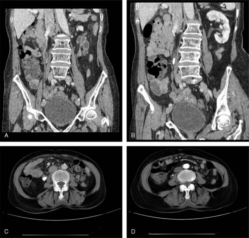
FIGURE 1.

Case 1. (A,B) MPR and (C,D) transverse scanning images display ureterostenosis of the right ureter at the level of the lower margin of the 4th lumbar vertebral body. (B,C) Dense stones and ureterectasia found at and above the place of ureterostenosis, respectively. (C,D) Dilated circuitous ovarian vein nearby that crosses over the ureter is displayed (white arrow indicates the ureter and black arrow indicates the ovarian vein). MPR = multiple planar reconstruction.
