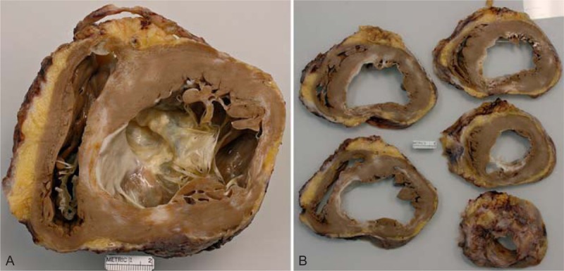
FIGURE 10.

Ischemic cardiomyopathy. Heart in a 61-year-old man who had a acute myocardial infarct at age 53 years followed by a coronary artery bypass grafting. By age 59 years, his left ventricular ejection fraction was as low as 15%. An intracardiac defibrillator had been inserted also at age 54 years. (a) View of the basal portion of the heart showing a portion of tricuspid valve and a good bit of the anterior mitral leaflet. A large transmural scar is present in the lateral wall between the 2 left ventricular papillary muscles and in the ventricular septum in apposition to the lateral wall infarct. The left ventricular cavity is greatly dilated. (b) View of the more caudal portions of the ventricular wall again showing the 2 infarcts extending almost to the apex of the heart.
