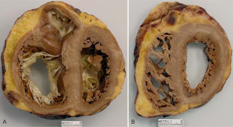
FIGURE 17.

Idiopathic dilated cardiomyopathy. Heart of a 68-year-old man who was found to have heart failure at age 50 years, and, at the time, also to have severe mitral regurgitation. Coronary angiogram showed the coronary arteries to be wide open. At age 50 years, the patient underwent repair of the mitral valve including the insertion of an annular ring. The mitral regurgitation was reduced from severe to mild, but the heart failure continued and progressed. A left ventricular assist device was inserted before the cardiac transplantation. (a) View of the basal portion of the heart showing dilatation of both right and left ventricular cavities. The tricuspid annulus is quite dilated. No lesions are noted in the ventricular walls although the ventricular septum is thicker than the left ventricular free wall. (b) View more caudal showing impressive pectinate muscles within the right ventricular cavity. Again, no myocardial lesions are noted. The quantity of subepicardial adipose tissue is considerably increased.
