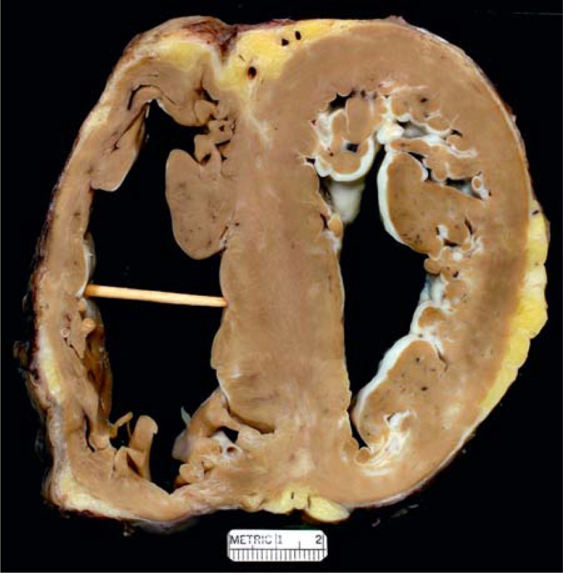
FIGURE 21.

Hypertrophic cardiomyopathy. View of the heart in a 68-year-old man who was known to have some form of heart disease since age 38 years. The problem during the 30 years was various degrees of heart failure which began progressing about age 55 years. In the 2 years before cardiac transplantation he had multiple hospitalizations for decompensated heart failure. He had an intracardiac defibrillator in place for several years. Because of the severity of the heart failure, a left ventricular assist device was inserted 7 months before cardiac transplantation. The heart failure severity, however, continued, and before heart transplantation the left ventricular ejection fraction was approximately 10%. Both cardiac ventricles are dilated, the right more than the left. The ventricular septum is thicker than the left ventricular free wall. The mural endocardium of the left ventricle is thickened by white fibrous tissue. A scar is present in the posterior portion of the left ventricular free wall and also in the posterior portion of the ventricular septum.
