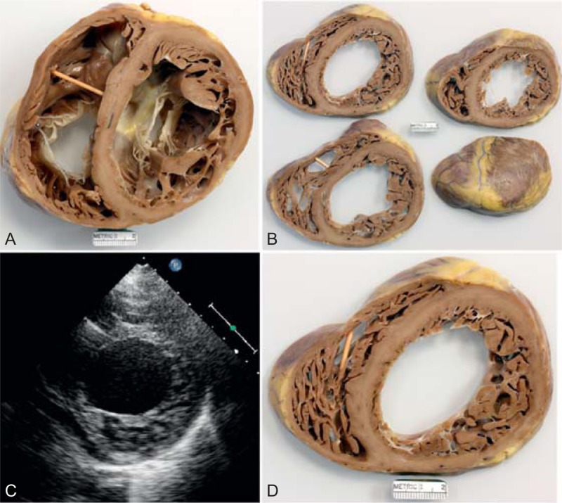
FIGURE 22.

Left ventricular non-compaction cardiomyopathy. Heart in a 49-year-old man who had been well until age 33 years when symptoms of heart failure appeared. At the time, echocardiogram showed both ventricles to be dilated and the left ventricular systolic function to be severely depressed. A defibrillator was inserted at age 39 years, and cardiac resynchronization therapy at age 43 years. He underwent ablation therapy for multiple episodes of ventricular tachycardia. His left ventricular ejection fraction just before cardiac transplantation was 15%. (a) View of both ventricles just caudal to the tricuspid and mitral valves. The thickness of the ventricular septum is much greater than that of the left ventricular free wall. The compacted portion of left ventricular free wall is much thinner than the non-compacted portion. Both ventricular cavities are greatly dilated. The left ventricular free wall posteriorly is focally scarred. (a) View of the basal portion of the heart exposing the tricuspid and mitral valves. Both ventricles are greatly dilated. The posterolateral wall is focally scarred. The compacted portion of left ventricular free wall is much thinner than the trabeculated portion. (b) Views of sections of the ventricular walls caudal to the view shown in the upper left. (c) Echocardiogram showing hypertrabeculation of the left ventricular free wall. (d) Section of the left ventricular free wall corresponding to the echocardiogram. (Reprinted with permission from Elsevier.45).
