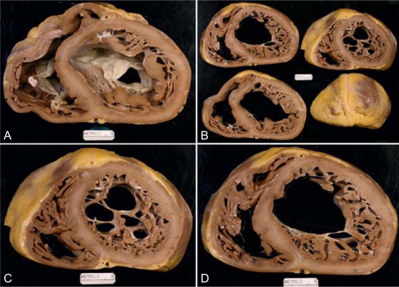
FIGURE 23.

Left ventricular non-compaction cardiomyopathy. Heart of a 36-year-old woman who was well until approximately age 35 years when she developed evidence of heart failure, which slowly but gradually progressed thereafter such that when she was aged 34 years her left ventricular ejection fraction had fallen to 20% and she was in chronic atrial fibrillation. She also developed runs of ventricular tachycardia for which a defibrillator was inserted. (a) View of the heart showing both tricuspid and mitral valves. Both ventricular cavities are enormously dilated, and the ventricular walls are free of foci of fibrosis and necrosis. (b) Views of the ventricles caudal to the view shown in upper left. (c) Close-up view of 1 of the slices in upper right. (d) View showing the hypertrabeculated or non-compacted portion of the left ventricular wall to be much thicker than the compacted portion. (Reprinted with permission from Elsevier.45).
