
FIGURE 2.
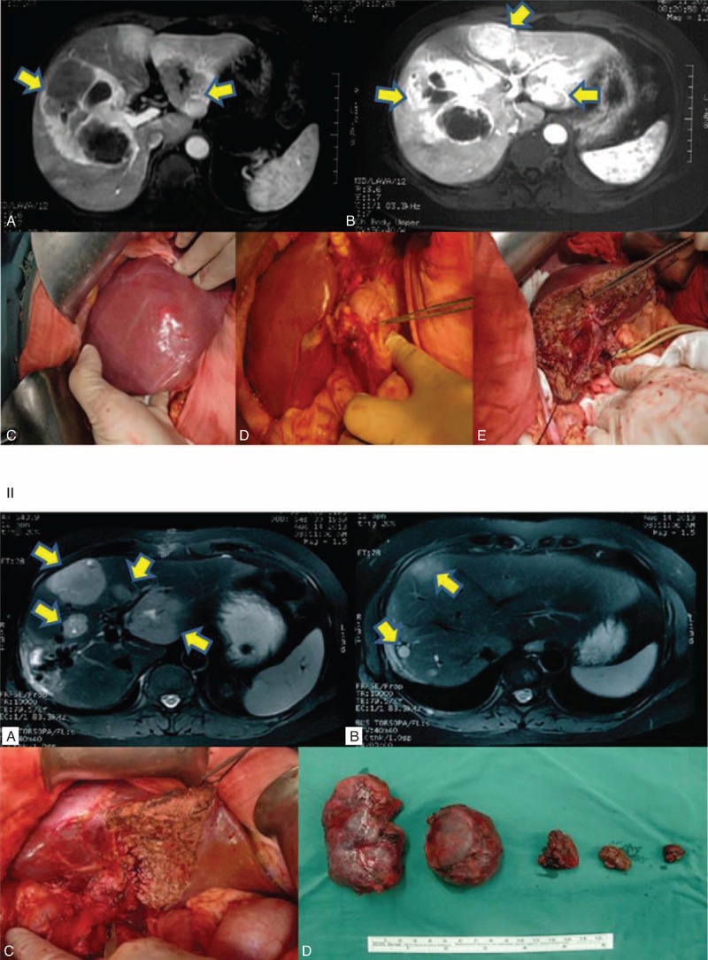
MRI and surgical data on 1 patient with G2-level NELM. A 52-year-old female patient was diagnosed with NELM with 6 liver neoplasms totaling 31 cm in size. Two separate surgical resections were performed. The first resection removed the largest tumor of 15 cm. The liver was allowed to regenerate to sufficient volume to sustain secondary resection. All 5 other neoplasms were removed during the secondary resection, which was performed 4 months after the first one. The patient was in good condition at the time of the preparation of this manuscript. Panel I, MRI images and key steps of first resection. (A & B) MRI images, arrows indicate locations of tumors; (C) a 15-cm tumor was found that covers sections of V, VI, VII, and VIII of liver; (D) primary tumor was found on the lesser curvature of the stomach; (E) remnant tissue of right lobe after resection. Panel II, MRI images and photos of second resection. (A & B) MRI images, arrows indicate locations of tumors; (C) remnant tissue after resection; (D) tumors resected from second operation. MRI = magnetic resonance imaging, NELM = neuroendocrine liver metastases