
FIGURE 3.
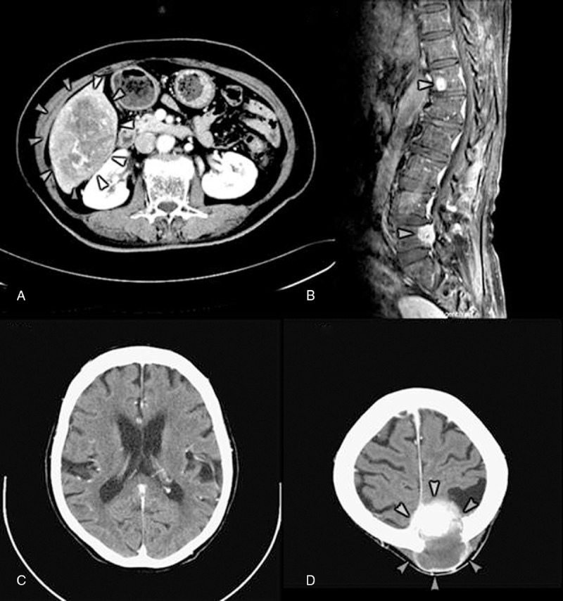
Images of case 2. (A) Abdominal CT with contrast shows a hypervascular tumor sized 11.0 × 7.5 × 9.1 cm over S5 and S6 of liver (arrowheads). (B) MRI of T-L spine (T2WI) shows degenerative spondylolisthesis at L4-L5 and multiple bony metastasis over T-L spine (T4, T6, T12 [arrowhead] and L5 [arrowhead]) with anterior epidural invasion and thecal sac compression at L5. (C) Brain CT with contrast shows small low densities involve bilateral basal ganglion, left thalamus, and left periventricular white matter, in favor of recent ischemic infarction. (D) Brain CT with contrast displays a mass lesion of 5.2 × 5.4 × 5.0 cm, manifesting hypervascularity, partial enhanced solid component and partial cystic change, involving left high parietal bone with skull destruction, epidural extension, and suspicious superior sagittal sinus invasion with compression of adjacent brain parenchyma (arrowheads). Left high parietal bone metastasis with epidural extension is indicated. CT = computed tomography, MRI = magnetic resonance imaging.